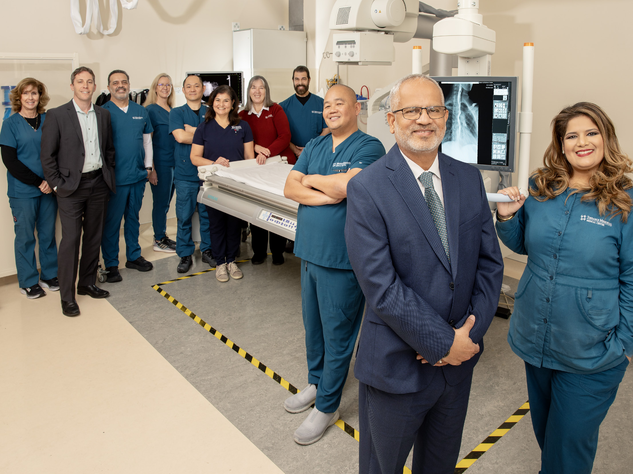
Under The Surface: Imaging Provides Information Critical to Diagnosis and Treatment
Struggling to breathe, the patient is rushed to the emergency department. The emergency physician orders a chest X-ray, which rules out pneumonia and other chest-related diseases. Next, the patient undergoes an ultrasound to check for a clot in his legs and a CT scan to look for clots in his chest. The CT scan reveals a pulmonary embolism — a clot in the arteries sending blood to the lungs. He then goes to the interventional radiology suite, where physicians pinpoint and remove his clot.
“At this point, the patient has undergone four modalities of imaging: X-ray, ultrasound, CT and interventional radiology,” notes Khalid Shariff, Torrance Memorial Medical Center’s director of imaging services. “Thanks to the skill of practitioners and advances in technology, the patient is able to go home the same day he experienced what was previously a fatal condition.”

